The University Medical Center Groningen and Kaer Labs announce entering into a research collaboration to evaluate NIR-II fluorescence for biological research and surgical guidance.
In the last 10 years, fluorescence imaging for surgical guidance has gained an unprecedented interest. The technique has been developed among others to help surgeons see tumors during the operation. Such a technique requires the use of a molecule with two key properties:
- the ability to bind specifically to the tumor that’s being removed
- the ability to be imaged, thanks to fluorescence properties
Specific instrumentation, namely intra operative fluorescent cameras, can then be used to reveal the presence of the molecule in the tissues and thus inform the surgeon about the position and extent of the tumor, right in the operating room and during the operation. Unfortunately, the type of fluorescence imaging currently used, in the NIR-I region, has limited performances in terms of depth penetration and resolution: small tumors or deep tumors can hardly be detected.
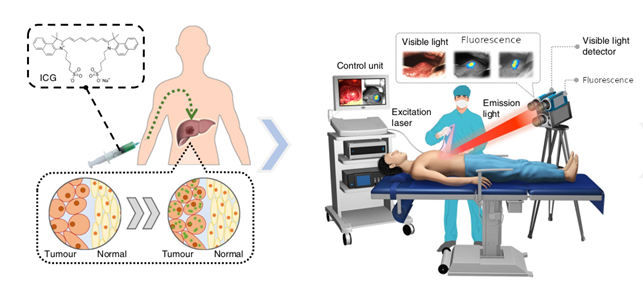
Principles of intra operative fluorescence guidance, modified from the illustration in the article from Hu et al, Nature Biomedical Engineering https://doi.org/10.1038/s41551-019-0494-0
Fluorescence imaging in the near-infrared I region (NIR-I, 700~950 nm) is indeed restricted by moderate signal–background ratios and low tissue penetration. Therefore, its clinical application is limited for obtaining detailed information on physiological or pathological processes, including vascular systems. Recently, second near-infrared (NIR-II, 900-1700 nm) fluorescence imaging technique has emerged, providing advantages in weak background noise, high spatial resolution, and deeper penetration depth. Because the quality of photon attenuation, tissue autofluorescence, and scattering are all significantly reduced when imaging at gradually longer wavelengths, NIR-II fluorescence imaging opens many exciting new imaging avenues for biological research compared to any other optical imaging modality.
The collaboration aims at assessing the performance of NIR-II fluorescence for different applications for biomedical research and experimental clinical guided surgery. The collaboration will start by assessing the possibility to use NIR-II fluorescence to visualize cetuximab-800CW in vitro, both on tissue slices and on surgical specimens just resected from patients injected with cetuximab-800CW. The results from both NIR-I and NIR-II fluorescence imaging techniques will be compared to see if the optical advantages of NIR-II imaging lead to the improvement of margin assessment.
UMCG and Kaer Labs also aim at using the NIR-II camera from Kaer Labs in vivo during surgical procedures. The system will be used to assess parathyroid gland perfusion in order to prevent hypoparathyroidism, a known and critical complication of thyroid surgery. The project may also include in vivo fluorescence-guided imaging for resection margin evaluation and detection of lymph node metastasis in different cancer types after injection of fluorescent markers like bevacizumab-800CW and cetuximab-800CW.
“Fluorescence is becoming increasingly important in a number of oncological surgeries, to guide surgeons during tumor resection or to help assess organ function”, says Schelto Kruijff, surgical oncologist and principal investigator from UMCG for the project. “We have seen major advances in the development and use of specific molecules that offer now more reliable information on tumor margins, for example. One of the key advantages of NIR-II, which could speed up its adoption, is the possibility to use the exact same workflow validated with NIR-I, as the instrumentation is similar in its use, and several NIR-I fluorescent agents are also NIR-II compatible. Yet it has the potential to significantly improve the detection performances and therefore the precision of the information provided. Although this is a preliminary work for the evaluation of NIR-II, we believe that such research projects are important in our effort to develop and validate the most relevant techniques for image guided surgery.”
“From the date of our publication in Nature Medicine, in 2011, where we showed that tumor-specific fluorescence imaging can increase visualization of solid tumors[1], paving the way for adequate margin assessment using optical imaging, the expectations from surgeons have been increasing”, says Prof. dr. Go Van Dam, surgeon oncologist and professor of Surgery at Groningen University, who published the first in-human application of targeted fluorescence. “There are unfortunately still major limitations that prevent the technique to be more rapidly and broadly adopted. Near infrared fluorescence, although it is already an improvement over visible fluorescence, is far from ideal in terms of optical performances to image biological tissue. The improvement of NIR-II on the quality of images could therefore be a great help to meet surgeons’ expectations and help them visualize the tumors more clearly. I am glad that the work around optical molecular imaging, that we initiated at UMCG, is brought forward by Prof. dr. S. Kruijff, with the example of this collaboration with the company Kaer Labs.”
 “We are very pleased by this project and this collaboration”, says Kaer Labs’ CEO, Pierre-Alix Dancer. “It is very important for us to put our NIR-II instrumentation in the hands of researchers like Prof. dr. Schelto Kruijff’s staff: they have the experience of evaluating technical progress and turning into surgically relevant practice. NIR-II can raise great hopes, but we want to identify precisely and as soon as possible the applications for which it will really make an impact for the benefits or biomedical research, and, later, of clinical practice.”
About Kaer Labs
Kaer Labs is a company specialized in the development and the sales of optical instrumentation for medical research and life sciences. It has developed the Kaer Imaging System for vivo fluorescence imaging in the NIR-I or NIR-II fluorescence, as well as a system for fast and easy 3D histology. The company was founded in 2018 in Nantes and has clients among academic institutions as well as pharmaceutical companies in France, Germany Belgium, Denmark, and also in Asia. Kaer Labs has several collaborations in Nantes (Imaging platforms MicroPICell / Nantes University and APEX / UMR703 INRAE/Oniris, and the laboratory RMeS / INSERM UMRS 1229 – Nantes University), in France (Imaging platforms: Optimal / University of Grenoble Alpes, LIOPA optical animal imaging platform / Paris University (UMS 3612 CNRS), Imaging and Cytometry Platform (PFIC), Gustave Roussy, Villejuif, and Molecular Chemistry Institute, Burgundy University (ICMUB) in Dijon; Research labs: team DiMABio, from the Fresnel Institute in Marseille; and the research and teaching institute IRCAD in Strasbourg) and in Europe (ICMI from VUB, Brussels, Belgium; Congenital Heart Disease surgical research unit, Dept of cardiac surgery, Rigshospitalet, Copenhagen University Hospital, Denmark; and the CRO Preclinics, Potsdam, Germany). Kaer Labs is a member of Atlanpole Biotherapies, It is supported by Atlanpole, IMT Atlantique, and Réseau Entreprendre Atlantique.